


Stroke
A condition that affects us all
The brain is the control center of the body. Although it only makes up 2% of our body weight, it consumes about 20% of our body’s energy supply. Around 1,100 liters of blood are pumped through it each day, supplying the brain with some 75 liters of oxygen and 115 grams of sugar. When a stroke interrupts the blood supply to specific areas of the organ, brain cells begin to die within a few minutes. For millennia, humans were powerless against this process.

Title page of Galen’s Opera Omnia, published in Venice in 1556
Source: Wellcome Library, London
Over 2,000 years ago, physicians described this condition in the Hippocratic Corpus and gave it the name apoplexia – a word that implies a sudden, violent blow. Treatment options were extremely limited: “It is impossible to cure a severe attack of apoplexy, and difficult to cure a mild one.” The celebrated Greek physician Galen therefore recommended a balanced diet in combination with running and sport – preventive measures that are just as applicable to reducing the risk of stroke today. Until the seventeenth century, it was assumed that strokes were caused by an imbalance in the mixture of blood, yellow bile, black bile, and phlegm – that is, the four humors described in the humoral theory that was developed in antiquity to explain the processes taking place in the human body.
It was not until 1658 that Johann Jacob Wepfer, a physician practicing in Schaffhausen, Switzerland, identified the root causes of stroke. Based on postmortem examinations of people who had died of the condition, Wepfer identified two forms of stroke that modern medicine still distinguishes between today.

Almost 85% of strokes are triggered by an “ischemic insult,” in which a blood clot blocks a vessel in the brain, interrupting the blood supply to certain areas of the organ. If, on the other hand, the condition is caused by a brain hemorrhage, physicians refer to it as hemorrhagic stroke. Given the fundamental differences in how these two forms are treated, it is vital to ascertain the type of stroke as quickly as possible.
Diagnosing stroke
For a long time, external symptoms such as signs of paralysis were all that physicians had to go on when it came to making a diagnosis. With the discovery of X-rays on November 8, 1895, however, they were able to gain unprecedented insights into the bodies of living patients. Many diseases could now be diagnosed significantly faster and much more reliably, although imaging the brain presented a particular challenge. Our brain is very well protected, surrounded as it is by the skull, meninges, and cerebrospinal fluid – and this protective housing severely limited the quality of diagnostic images in the early days. The development of special devices and examination methods gradually paved the way for a clearer view of the brain and its vascular system, although the laborious and often protracted examinations were extremely unpleasant for patients. In the 1970s, the development of computed tomography (CT) led to a breakthrough in stroke diagnostics: For the first time, physicians could quickly and reliably discern whether they were dealing with a circulatory problem or a cerebral hemorrhage. The emergence of magnetic resonance imaging (MRI) and Doppler and duplex sonography allowed further information to be obtained about a stroke. But what use is the most accurate diagnosis unless it is accompanied by suitable treatment options? Until the mid-1990s, there was still no effective way of treating acute cases of stroke.
Milestones in cerebral angiography

1927
Antonio Egas Moniz uses X-ray technology to produce the first image of the cerebral arteries in a living person.

1931
The Lysholm skull unit is the first specialized device for imaging the brain using contrast agents in the history of Siemens Healthineers. It remains the standard tool for complex cranial imaging and angiography of the brain for three decades.

1956
The cerebral cassette changer for serial angiography from Siemens is specially tailored to the serial imaging of cerebral blood vessels. Multiple X-ray images taken in quick succession allow the physician to observe where circulation is obstructed, for example, and what is causing the problem.

1975
Siemens launches its first CT scanner: the SIRETOM cranial scanner.

1977
The Angioskop is the first device from Siemens to feature a C-arm that can be moved freely around the patient in every direction – albeit still by hand at this stage.By virtue of its construction, the device is also very well suited to cerebral angiography.

1983
Siemens presents MAGNETOM, its first commercial MRI system.

1990
The Q2000 color Doppler system allows not only the finest vessels but also arteriosclerosis and blood flow to be visualized without a contrast agent. It is still not possible for physicians to look inside a patient’s brain, however, as is stated in the press release announcing the launch of the system.

2005
SOMATOM Definition is the world’s first dual-source scanner and operates with two X-ray tubes at the same time. Thanks to its high speed and accuracy, SOMATOM Definition is also excellently suited to diagnosing suspected stroke patients in an acute care setting.
2006
Siemens presents the new MRI application Syngo SWI. Susceptibility-weighted imaging (SWI) allows physicians to identify even the tiniest brain hemorrhages and diagnose an imminent stroke faster than before.
2007
Siemens develops Syngo ASL in collaboration with the University of Pennsylvania. Arterial spin labeling (ASL) is an MRI technique that uses the water in arterial blood as an endogenous contrast agent. It offers insights into the circulation and functional physiology of the brain and is therefore also of interest for stroke evaluation.

2019
With the SOMATOM On.site mobile head scanner from Siemens Healthineers, physicians can perform CT scans directly at a patient’s bedside in the intensive care unit (ICU). This dispenses with the time-consuming and labor-intensive process of transporting the patient from the ICU to the radiology department.
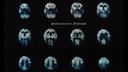
Today, computed tomography (CT) can produce high-resolution 3D images that allow physicians to make a detailed assessment of the skull, the soft tissues of the brain, and the structures of the cerebral vessels (Fig. 1). CT is routinely used in stroke diagnostics and can deliver automated results for the areas of the brain affected by the stroke (Fig. 2). Moreover, functional imaging helps physicians discern which areas of the brain can still be saved (Fig. 3) and is able to distinguish between active and inactive brain hemorrhages (Fig. 4) following surgery.

If less than 4.5 hours have elapsed since a stroke began, an anatomical MRI image will not usually show the accompanying edema (see ellipse in right image). On the other hand, cytotoxic edema – a condition caused by the death of neurons in the event of a stroke – can be imaged very effectively at an early stage (see ellipse in left image) using a technique known as diffusion-weighted magnetic resonance imaging. This can now be performed using devices such as MAGNETOM Vida and is an effective way for physicians to narrow down the time at which a stroke began.
1927
Antonio Egas Moniz uses X-ray technology to produce the first image of the cerebral arteries in a living person.
Clearing the blockage
In the 1970s, research by the Belgian molecular biologist Désiré Collen marked the starting point for the development of a medicinal treatment for ischemic stroke. In his search for a way to dissolve blood clots, Collen developed a substance known as alteplase recombinant tissue plasminogen activator (rt-PA), which can dismantle the underlying framework of the clot (or “thrombus”) and therefore break it down. Since the mid-1990s, this drug has been used for the acute treatment of stroke in lysis therapy (from the Greek lúsis, “loosening”). Some 50–60% of vascular occlusions can be eliminated in this way, thereby restoring the blood supply to the affected area of the brain. Lysis therapy is not suitable for every patient, however, and must be administered within a 4.5-hour window after stroke onset.
Thanks to pioneering work in the field of interventional radiology by physicians such as Werner Forssmann, Charles Dotter, and Andreas Grüntzig, lysis therapy is no longer the only treatment option available. Since approximately 2008, it has also been possible to treat vascular occlusions using mechanical thrombectomy, a technique in which physicians pass a catheter into the groin and through the carotid artery in order to reach a blood clot that is blocking a cerebral blood vessel. Using X-ray technology to monitor the procedure, the physicians can then remove the clot using a “stent retriever” (a basket-like wire mesh that travels inside the catheter) or a special aspiration catheter (a sort of tiny vacuum cleaner). Mechanical thrombectomy gives physicians a window of 24 hours between the onset and treatment of stroke, and therefore more time than is the case with lysis. Despite this, and although the two treatments can also be combined, the maxim “time is brain” always applies. The sooner a patient is treated following the onset of symptoms, the greater chance they have of avoiding lasting impairment.
Milestones in catheter therapy

1929
The young intern Werner Forssmann performs the world’s first cardiac catheter examination. In a daring self-experiment, he inserts a rubber tube into his heart via a venous access point, thereby proving that cardiac catheter examinations are possible.
Read the full story here.
Source: Forssmann, Werner: “Die Sondierung des rechten Herzens” (“Probing of the right heart”), Klinische Wochenschrift, No. 45, 1929

1963
While performing an angiogram on a patient with a narrowed renal artery, Charles Dotter accidentally dislodges the blockage with the catheter, allowing the blood to flow freely again. From this point onward, Dotter dedicates his entire career to “catheter therapy” and to his goal of treating patients with a catheter instead of a scalpel whenever possible.
Find out more!
Source: Maria Schlumpf-Walker

1977
Andreas Grüntzig carries out the first percutaneous transluminal coronary angioplasty (PTCA) using a self-built balloon catheter, thereby avoiding the need for bypass surgery on his patient – who was at serious risk of a heart attack.
Source: Maria Schlumpf-Walker

1990
The Italian neurosurgeon Guido Guglielmi, MD, carries out the first intervention in which a Guglielmi Detachable Coil – a platinum coil – is placed in a cerebral blood vessel in order to seal an aneurysm. This can prevent the aneurysm from bursting and causing a cerebral hemorrhage.
2005
The hybrid solution MIYABI Angio-CT combines a rail-mounted SOMATOM Sensation CT scanner with an AXIOM Artis angiography system, allowing the delivery of improved care for emergency patients. First, a complete vascular study of the brain can be carried out using the CT scanner, which can then be moved to the neighboring room. Should catheterization be necessary, the angiography system is standing by and can be moved into place immediately.
2008
For the first time, thrombectomies are performed using completely unfolded intracranial stents (stent retrievers).
Courtesy: M. Psychogios, MD, Head of Neuroradiology, University Hospital Basel, Switzerland

2021
syngo DynaCT Multiphase makes it possible to visualize cerebral collateral vessels with time-resolved syngo DynaCT, depicting 10 different time points within a period of 60 seconds.
1929
The young intern Werner Forssmann performs the world’s first cardiac catheter examination. In a daring self-experiment, he inserts a rubber tube into his heart via a venous access point, thereby proving that cardiac catheter examinations are possible.
Read the full story here.
Source: Forssmann, Werner: “Die Sondierung des rechten Herzens” (“Probing of the right heart”), Klinische Wochenschrift, No. 45, 1929
Over the last 30 years, rapid advances in diagnosis and treatment options have played a key part in reducing the mortality rate after stroke. Swift treatment and optimized workflows can considerably reduce the impact of the condition, but it is still the second most common cause of death worldwide. Therefore work will also continue with a view to improving treatment options and promoting preventive measures, in the future. Mechanical thrombectomy in particular is one promising approach. Although there are still relatively few experts who can perform this procedure, stroke patients around the world could benefit from the technique thanks to artificial intelligence and robot-assisted technology in the future.
Expert for History Communication and Historian at the Siemens Healthineers Historical Institute