


Siemens Healthineers MedMuseum
Innovations
Innovations transform the world of medicine
Since 1847, we have shaped the development of medical technology through our numerous innovations. Join us on a journey through the history of Siemens Healthineers with a selection of our most important innovations from 1847-2010.

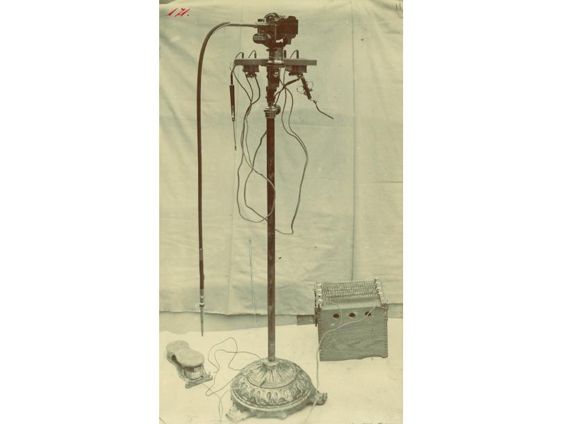
1847
The slide inductor for electrotherapy was the first medical device manufactured by Siemens & Halske. It was marketed successfully for decades.
1891
The first electric dental drill in Germany, which was manufactured by RGS, gradually replaced the previous foot-operated models.

1896
When X-rays were discovered, both Siemens & Halske, and Reiniger, Gebbert & Schall immediately realized their potential and soon began producing X-ray equipment.

1909
Friedrich Dessauer’s “Blitzapparat” made scans taking several minutes a thing of the past. With an exposure time of just a few milliseconds, it was the first device that could capture well-defined images of the heart.
1910
The Pantostat was one of the first multifunctional devices for use in electromedicine. From a four-cell galvanic bath to a vibration massage, it had numerous applications and was a bestseller all over the world until the 1970s.

1913
Based on telephone technology, the Phonophor was the first hearing aid launched by Siemens & Halske.

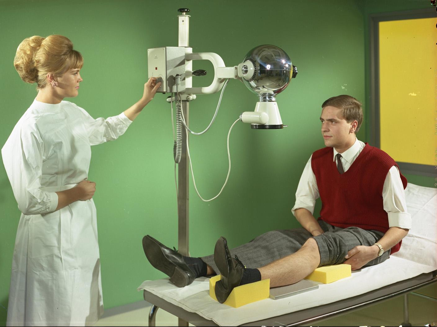
1916
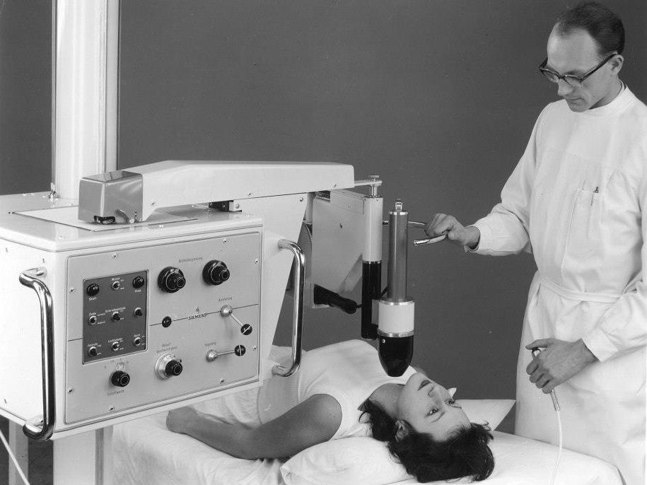
The Symmetry apparatus was the first X-ray generator specifically developed for deep treatment.
1933
The Pantix was the first X-ray tube with a disk-shaped rotating anode. This design was subsequently adopted by all tube manufacturers and forms the basis of rotating anode X-ray tubes to this day.
1934
The X-ray Sphere combined a transformer and an X-ray tube within a single unit, making it economical, easy to transport and operate, and suitable for direct connection to the electricity supply.
1941
The effervescent Clinitest tablet contained various reagents and could be mixed with urine to provide a quick indication of blood sugar level based on color changes. It laid the foundation for modern point-of-care diagnostics.
1949
Quick and cost-effective screening became possible thanks to the Seriomat, particularly with regard to the early detection of tuberculosis. The device saw worldwide use during the 1950s and 1960s.
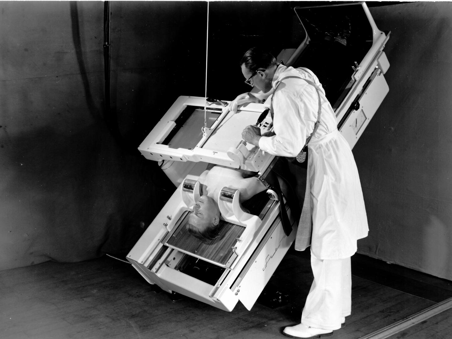
1950
The Betratron electron accelerator allowed the effective irradiation of tumors deep within the body for the first time, marking a decisive breakthrough in radiation therapy. |

1951
The Mingograph was the first direct-writing electrocardiograph to plot cardiac activity graphs directly onto paper used a jet of ink instead of onto film. This enabled much more accurate recordings and faster evaluations than in the past.

1951
The Angiograph was the first system to allow a catheter to be observed on a fluorescent screen as it passed through the blood vessels and into the heart.
1953
The basic framework of the SIRESKOP was a completely redeveloped patient table with numerous adjustment aids, such as electric motors and magnetic brakes. The device was seen as a breakthrough in both imaging and operating technology and remained in production for a number of decades.
1953
Inge Edler and Carl Hellmuth Hertz successfully used a Siemens materials testing device optimized for medical use to perform the first noninvasive visualization of cardiac function using ultrasonic waves. The technique of echocardiography was born. |
1956
Clinistix were the first ever test strips and simultaneously introduced a new method known as “dip-and-read” diagnostics. These reagent-containing test strips can be used to determine the blood sugar level in bodily fluids. Compared with previous tests, the new method proved to be easier, faster, and more reliable.

1957
The AutoAnalyzer was the first step toward modern laboratory automation. The key innovation in this system was that the samples were separated by air bubbles and flowed through the device continuously. |
1957
The X-ray image intensifier generated fluoroscopic images of the body that could be seen by daylight. This made it possible, for the first time, for physicians to check a patient by X-ray during surgery without lowering the lights. |
1958
The Nucleograph was the first nuclear medicine scanner from Siemens. It produced a scintigram showing the distribution of radioactive substances injected into the body and therefore allowed the visualization of metabolic processes. |

1958
The engineer Rune Elmqvist revolutionized cardiology when he developed the world´s first fully implantable cardiac pacemaker for Elema-Schönander, which later became Siemens-Elema.

1960
As the first commercially available, fully rotational linear accelerator for radiotherapy, the Clinac 6 from Varian delivered proof that linear accelerators could be used to treat cancerous tumors.

1967
With the Vidoson, Siemens developed the world’s first ultrasound system to allow real-time observation of movements inside the body. |

1968
The Clinac 4 from Varian was the first medical linear accelerator based on standing wave technology, leading to a reduction in size, costs, and the complexity of operation.
1965
An X-ray examination table for the most stringent requirements, with a combined image intensifier and television screen. The Siregraph was one of the most successful product lines over the next 35 years and even featured on a German 60 Pfennig stamp in 1978.

1972
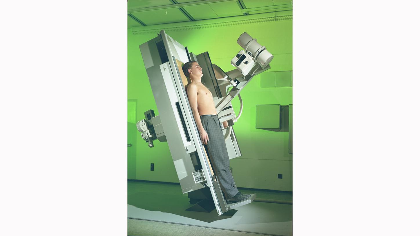
The Mammomat was the first Siemens system designed specifically for examining the female breast.
1975
Siretom was the first computed tomography (CT) scanner from Siemens. The head scanner imaged the brain in slices and was used to examine suspected brain diseases, for example, or for surgical planning.

1977
Somatom was the first whole body CT scanner from Siemens.

1983
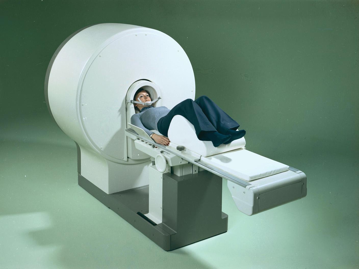
MAGNETOM was the first commercial magnetic resonance imaging (MRI) scanner from Siemens. This new technology allowed the slice-by-slice imaging of fine structures in soft tissue.

1983
The ACUSON 128 was far superior to all other devices of its era. For the first time, the ultrasound image was computer-generated and optimized using software. The two-dimensional image could be enlarged and viewed in real time.

1990
SOMATOM Plus-S was the first spiral CT scanner and offered seamless scanning at a higher speed than previous CT scanners. As result, movements inside the body no longer caused a reduction in image quality.
1990
Varian’s multileaf collimator could be used to dynamically adapt the treatment beam to the shape of the tumor with the help of 52 separate leaves.

1998
With SOMATOM Volume Zoom, Siemens developed a system with multislice technology that recorded several slices in a single rotation, reducing scanning times considerably.

1999
From X-ray systems to MRI scanners, the syngo image-processing software provided a common user interface for Siemens imaging systems.

2000
Biograph was the first system from Siemens to bring together positron emission tomography (PET) and computed tomography (CT) in a single device.
2003
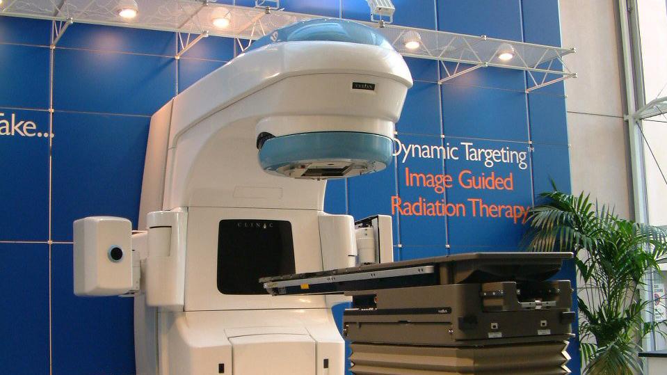
Varian’s Dynamic Targeting Image Guided Radiation Therapy (IGRT) technology tracked the tumor’s movement using the On-Board Imager (OBI). The OBI’s built-in cone-beam CT mode allowed clear identification of the tumor position even in complex cases.
2005
SOMATOM Definition operated using two X-ray tubes at the same time and could record an image of the entire heart in just 0.083 seconds.

2008
Artis zeego was the first multi-axis system with robotic technology in which the C-arm is synchronized with the movement of the table.

2010
Biograph mMR was the first system to allow simultaneous examination using magnetic resonance imaging (MRI) and positron emission tomography (PET).
2010
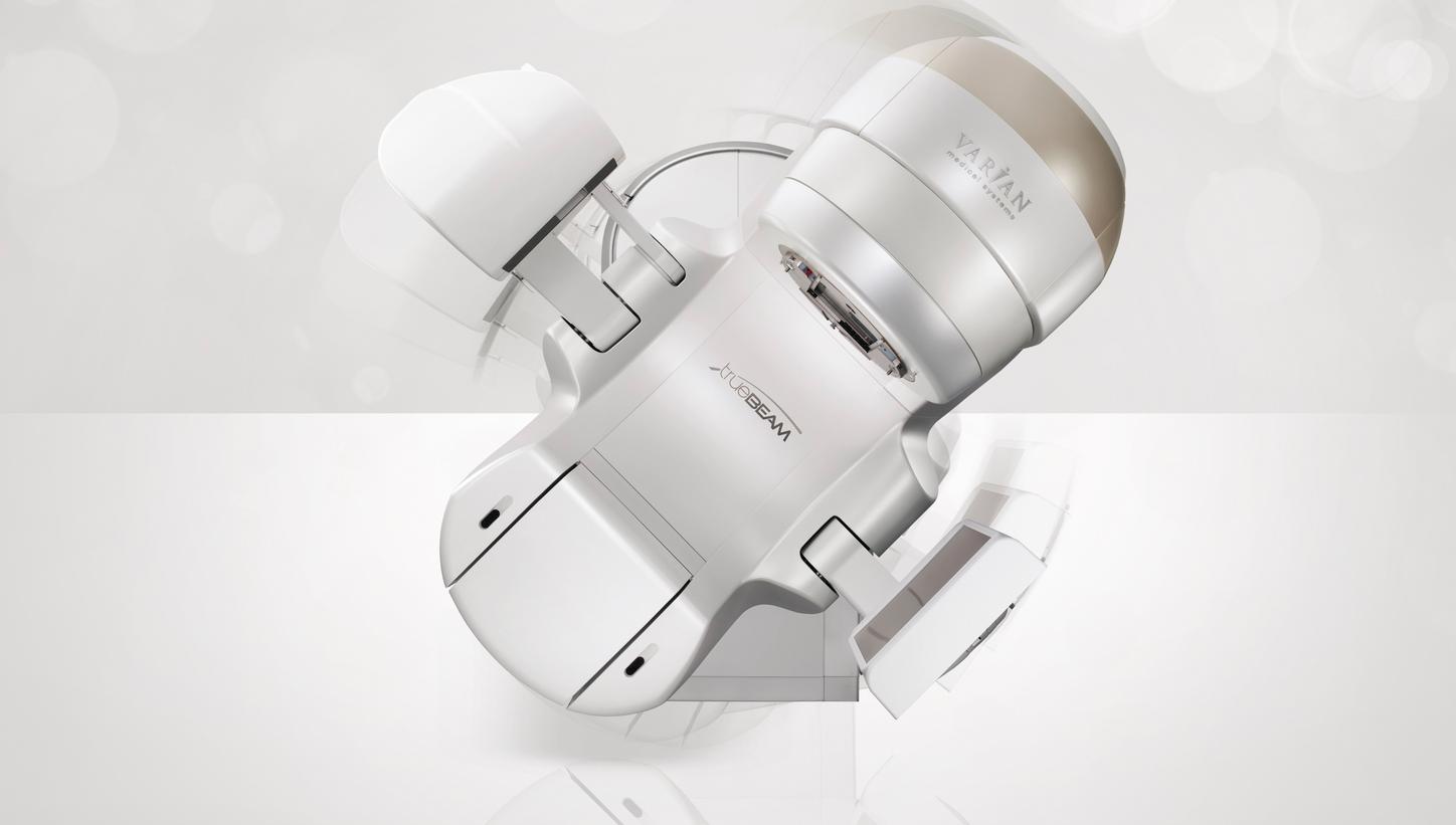
The TrueBeam linear accelerator dynamically synchronized all the latest technologies from the fields of imaging, patient positioning, and movement compensation. This approach significantly reduced the number of steps arising during treatment, and the desired radiation dose could be administered up to four times faster than with other linear accelerators.

2013
During image generation, the xSPECT technology of Symbia Intevo™ could take account of all SPECT and CT data in order to produce tomograms with high accuracy and resolution.

2017
Biograph Vision offers a new level of precision in PET/CT imaging. Biograph Visions leverages the groundbreaking technical performance to reveal the bigger picture - enabling users to more accurately and confidently diagnose, treat, monitor, and research disease.

2017
MAGNETOM Terra was the first 7-tesla system for clinical diagnostics. At the time of its launch, its actively shielded magnet was the lightest 7-tesla whole-body magnet in the world, weighing in at around 17 tonnes.

2017
With Halcyon, Varian revolutionized the radiation therapy process in many respects: Compared with conventional linear accelerators, the system consumed less than half the energy; the gantry rotated up to four times faster; the system’s self-shielding meant that two thirds less concrete was needed to build the bunker; and the operating personnel only needed to carry out nine instead of over 30 process steps from the start to the end of treatment.

2018
Even in patients with a high body mass index, ACUSON Sequoia delivers diagnostically relevant images from deeper regions of the body.

2019
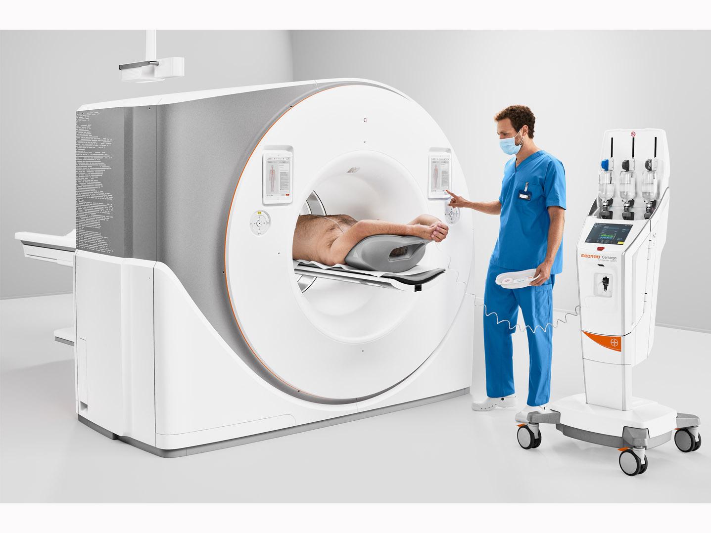
With a video camera in the gantry and an 82 cm wide bore, the high-end CT scanner SOMATOM X.cite allows constant contact between the patient and the medical staff, making examinations much less stressful for patients.
2019
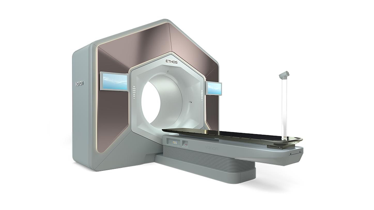
Varian Ethos uses artificial intelligence and machine learning to adapt the entire treatment process to the individual patient automatically — from initial planning to treatment monitoring. The system has such an intuitive design that it can be used in almost any hospital without the need for additional operating personnel with special training.

2020
MAGNETOM Free.Max was the world’s first MRI scanner with an 80-centimeter bore.
2022
NAEOTOM Alpha® was the first photon-counting CT scanner and paves the way for even higher-resolution images, allowing even fine details to be visualized.
1847
The slide inductor for electrotherapy was the first medical device manufactured by Siemens & Halske. It was marketed successfully for decades.