


Siemens Healthineers MedMuseum
Discover (hi)stories
- “Oh, wow! What are they doing there?”

“Oh, wow! What are they doing there?”
Pioneers working on new technologies often face unusual challenges. In the late 1990s, a team with the physicist Thomas Brunner presented an idea for a new technique that could produce computed tomography-like images directly using the angiography system in an interventional radiology room. The reactions ranged from skepticism to astonishment. “Oh, wow! What are they doing there?” is how Brunner summarizes the surprised response of many colleagues from the angiography systems division. At first, the team working on the development of computed tomography (CT) believed this project had poor chances of success. In their view, the limitations inherent in the structure of angiography systems probably made it impossible to produce usable CT-like images. “The structure of an angiography system isn’t really suitable for CT imaging,” says Brunner. “There are several limiting circumstances with a C-arm system. For one, the detector of an angiography system isn’t designed for CT imaging — and the C-arm’s maximum rotation speed is much lower than that of CT scanners.”
Interventional radiology
The term “interventional radiology” refers to a range of procedures, normally minimally invasive, that are performed with the aid of imaging systems.
Despite the seemingly poor chances of success, the team received considerable support from the outset. Management was in favor of developing this unusual technology, and colleagues in basic research and computed tomography were on hand to provide advice and practical assistance. There were some particularly unusual challenges in store for these teams. “For them, the process of working together on our idea was uncharted territory,” Brunner recalls over 20 years later. “It was very, very difficult to integrate our technology’s requirements into an angiography system.” In mid-2004, after around five years of research into various different prototypes, the technology had reached maturity and was given the name syngo DynaCT. This was a special moment for the development team and for the division, as Brunner recalls: “To see everything working as planned and that the first diagnostic images delivered the desired level of quality — that was such an amazing moment for us after overcoming a whole series of challenges.”

Head scan with syngo DynaCT from 2004
“They couldn’t believe what they were seeing.”
In November 2004, syngo DynaCT was presented to a wider audience for the first time at the Annual Meeting of the Radiological Society of North America (RSNA) in Chicago. In 2005, Siemens Healthineers (which was known as Siemens Medical Solutions in those days) presented AXIOM Artis, the first angiography system with DynaCT technology, at RSNA. Product manager Michael Martens still vividly recalls that era: “It was really astonishing to see how many people had gathered at our exhibition stand to catch a glimpse of our new system. It was a biplane system, which is a configuration typically used by our customers in neuroradiology. syngo DynaCT allowed those customers to witness CT-like imaging that could visualize things like bleeding in the brain or implanted stents for the first time.” The first syngo DynaCT images, which Siemens Healthineers exhibited at the RSNA Annual Meeting, were the first CT-like images produced using an interventional imaging system. “Many customers from different countries came to have a look. Some genuinely said the images looked like they’d been captured with their CT scanners, while other customers couldn’t really believe what they were seeing.” Some were initially skeptical, but they were won over to this innovative technology thanks to its ability to perform diagnostic imaging using an interventional system. “It makes me very proud as product manager that all the thoughts and ideas we had during development and testing turned into something that has had a significant impact on clinical practice and continues to benefit many patients to this day.”

AXIOM Artis, the first angiography system with syngo DynaCT technology
Nowadays, we can look back on numerous milestones, both small and large, in the history of syngo DynaCT technology. “We have many customers who contributed novel ideas, thoughts, and applications to our further development work,” says Martens. The first generation of the technology was optimized exclusively for visualizing bleeding in the brain. This remains a key application of syngo DynaCT to this day but has been joined by numerous other applications over the years. For example, shortly after the presentation in Nuremberg (Germany) and Chiba (Japan), syngo DynaCT was used to visualize for the first time tumors in the liver and kidneys, as well as their vascular supply. This was followed by the first three-dimensional image of a heart in 2007 and the first image of the blood volume in the brain in 2009. Then, in 2012, syngo DynaCT took the resolution of soft tissue to the next level in conjunction with the new HDR detector. In the latest generation, an acquisition of DynaCT can be now as fast as 2.5 seconds. Brunner is still working on the development of syngo DynaCT today. He smiles as he reflects on the technology’s 20th anniversary: “I think we’ve demonstrated that all the effort was worthwhile — and that the journey is far from over.”

Heart scan with syngo DynaCT Cardiac, 2007

Scan of the abdomen with syngo DynaCT Large Volume in 2008

Visualization of relative blood volume in the brain with syngo DynaPBV Neuro, 2009
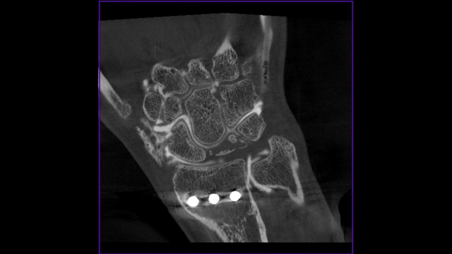
Scan of a triangular fibrocartilage complex on the wrist with syngo DynaCT Micro, 2012

Lung scan with syngo DynaCT High Speed in 2022

Visualization of the vascular tree during prostatic artery embolization with syngo DynaCT, 2023
Heart scan with syngo DynaCT Cardiac, 2007
Technikjournalist und Autor im Siemens Healthineers Historical Institute