


Siemens Healthineers MedMuseum
Discover (hi)stories
The green patient in the wooden shed
The green patient in the wooden shed
How the story of magnetic resonance imaging at Siemens Healthineers began.
In the mid-1970s, word got out about a fascinating new technology that could produce images of the inside of the human body using magnets and radio-frequency technology. The method, which today is commonly known as magnetic resonance imaging, was initially only used in universities and limited to imaging small parts of the body, such as fingers. In 1977, Siemens made the decision to develop a whole-body system for hospitals and radiology practices—a task that the development team solved in some creative ways.
When work on the first prototype began in February 1978, physicist Arnulf Oppelt and his team faced some unusual challenges. Not least because the development of a magnetic resonance (MRI) scanner differs considerably from the design of other medical engineering systems, such as X-ray machines or CT scanners. An MRI scanner works with magnetic fields that are a thousand times stronger than that of the Earth. Even minute influences in the vicinity of the scanner, such as passing automobiles or other metallic objects, caused interference in the technology available at that time and detrimentally affected the quality of the images.
In order to avoid this interference with the magnetic field, Siemens built a wooden shed devoid of magnetic parts on the grounds of the central research laboratory. Not one iron nail was used in its construction!

By mid-1979, the first wooden shed prototype stood and was ready for operation, but the team was still having problems with interference: The frequency of magnetic resonance is in the radio wave range and so signals from shortwave radio stations also affected the image quality. The problem could in fact have been easily solved with a filter, which the Siemens Energy division had in its portfolio; but weeks, even months, would pass before one could be delivered. The noise was still in the images—and November 1979 was steadily approaching. Oppelt and physicist Wilfried Loeffler, whose main task was programming the software, were expected to provide reproducible results before the budget review the following year. And this meant: an image.
The bell pepper had everyone excited!
"So I said to Wilfried Loeffler: No problem," Arnulf Oppelt recounted over 40 years later. "We'll just scan for a very long time, again and again, and the interference will be averaged out." For an acquisition time of several hours, the two of them needed a measurement object that remained still, had a structure, and that could be cut open to show that the image really does visualize the internal structure. "We needed something that contains water," Loeffler explained. So they both drove to a local produce store and purchased a large, juicy green bell pepper. "We placed it in the prototype and said: OK, now let's set the thing to scan for two hours." By the evening, all the data had been collected. Oppelt and Loeffler planned to calculate the images the next morning and went home. "The next day we reconstructed the image and got quite excited," Oppelt recounted. "We then showed it to our bosses and they were even more excited. The bell pepper had got everyone excited!"

The MR image of the bell pepper showing the capture date February 14, 1980. In reality, the image was taken in November 1979; however, a minor image error had to be eliminated soo the data were reconstructed in 1980.
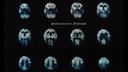
It became immediately obvious that Oppelt and Loeffler had to continue with their work. Just four months later, the two of them were able to present the next major development milestone: an MR image of the head of their boss Alexander Ganssen.

From February 1983, Siemens successfully tested an advanced version of the prototype at the Hannover Medical School under clinical conditions for suitability in daily routine.

In August 1983, after an installation period of just under three months, the first commercial MRI system in the history of Siemens Healthineers was commissioned at the Mallinckrodt Institute of Radiology in St. Louis (USA): the MAGNETOM GBS 1.

The valuable role that magnetic resonance imaging would play in diagnosis had already become apparent: At no time in the past had soft tissue such as that of the human brain been visualized with such detail and contrast. The experts were quick to agree that a great future lay ahead for this technology. Today—40 years and many technological milestones later—MRI is one of the most important diagnostic imaging methods available to medicine.
Read more!
The history of magnetic resonance imaging at Siemens Healthineers
(pdf, 6890.64 KB)Technology journalist and author at the Siemens Healthineers Historical Institute