


Siemens Healthineers MedMuseum
Discover (hi)stories
- Organs in one go
Organs in one go
After completing his physics degree and working at Harvard University for two years, Anger applied to the University of California, Berkeley with a view to working at the laboratory of physics Nobel laureate Ernest O. Lawrence. In 1946, Anger embarked on his first day of work at the Lawrence Radiation Laboratory. Besides his tasks as an engineer, Anger frequently had the chance to implement his own ideas and to experiment with new approaches at the lab. Among other things, he invented the well counter in 1950. This device is now one of the most commonly used instruments in nuclear medicine laboratories. A year later, Anger read about the invention of a new imaging technique in nuclear medicine that allowed human metabolism to be visualized as a line pattern.
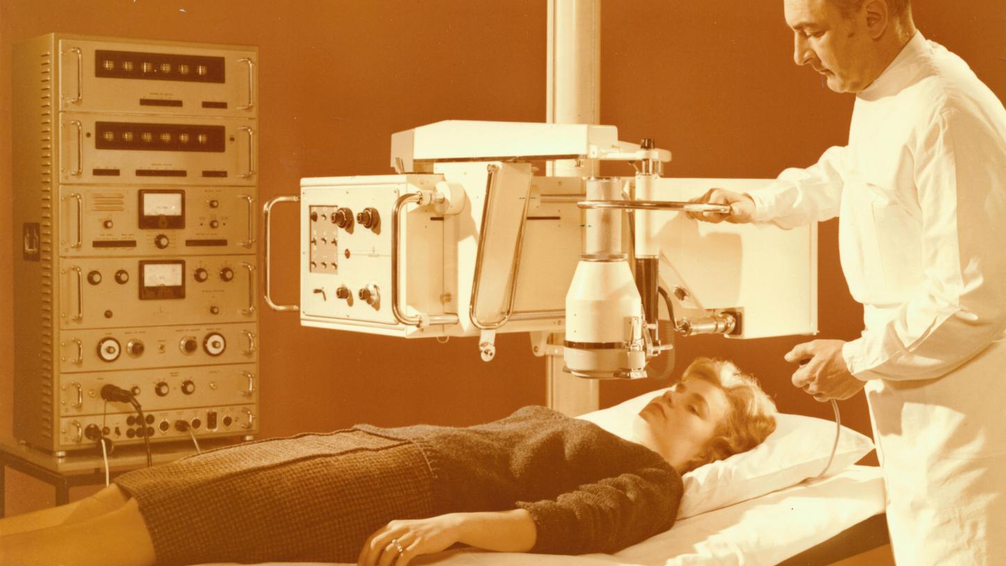
In the mid-20th century, rectilinear scanners such as the Nucleograph from Siemens allowed human metabolism to be visualized for the first time.
What is molecular imaging?
Metabolic processes inside the human body can be visualized using equipment based on the principles of nuclear medicine. In a technique known as “molecular imaging,” patients are given a substance known as a tracer before the scan. This is generally a type of weakly radioactively labeled glucose. The tracer participates in metabolic processes without disturbing them while at the same time emitting tiny doses of radiation over the course of a few hours.
Among other equipment, nuclear medicine examinations can be carried out using “gamma cameras.” These instruments contain multiple very wide measuring heads that convert the gamma rays from the tracer into electrical signals. Inside each measuring head, the radiation first collides with crystals, where it produces scintillations (flashes of light, from Latin “scintilla”, meaning spark). Then, photodiodes or photomultipliers convert this light into electrical signals, which can be amplified and relayed in order to generate the image.
The rectilinear scanner developed by the medical physicist Benedict Cassen scanned organs point by point by moving a detector head mechanically across the patient. This small, very slow-moving detector had several shortcomings. These included the fact that the line patterns had a very low resolution, the measurement process could take over half an hour, and the diagnostic image was not a real two-dimensional representation of metabolic processes. Anger wanted to develop a device that could image a whole organ at once, just as a camera captures and depicts everything in its field of view simultaneously.
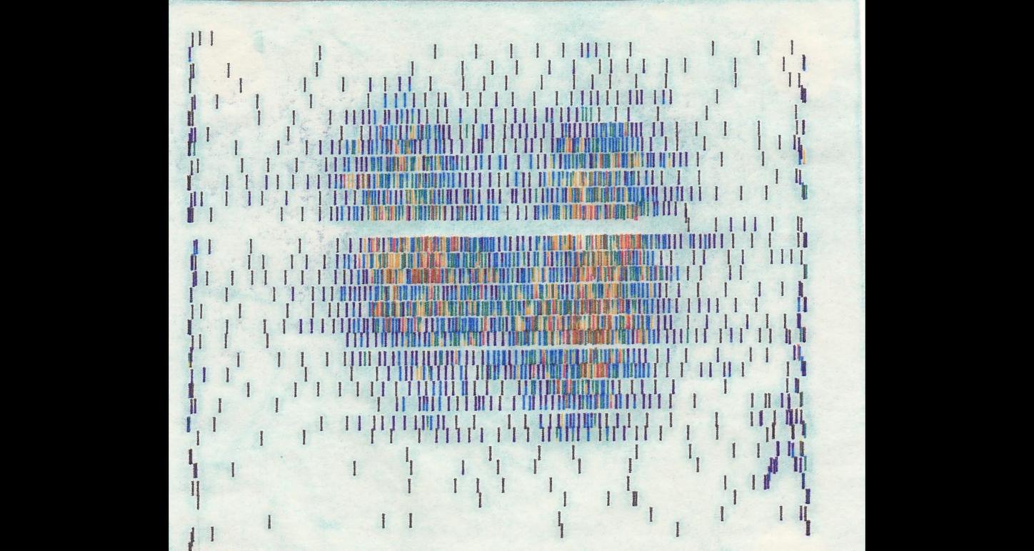
Scan of the metabolism of a thyroid gland as a line diagram (scintigram) with a rectilinear scanner from Siemens.
Accordingly, Anger’s first prototype from 1952 was essentially built like a pinhole camera: A collimator guided the rays onto a crystal with a photographic plate placed on it in order to record all the gamma flashes as points with approximate spatial accuracy. This method proved to be insufficiently sensitive, however, with a scan taking over an hour. Following numerous improvements, Anger’s nuclear medicine camera of 1956 worked on a basic principle so sophisticated that it would be decades before it could be significantly improved upon. Specifically, the camera contained a large, single crystal with a diameter of about 10 centimeters. Coupled to the crystal were seven photomultipliers, which converted the gamma flashes into electrical signals. The camera’s electronics were wired up in such a way that they could calculate the position of the gamma flashes based on a complex system of logic. To this day, this method of calculation is still referred to as Anger logic.
In 1958, Anger presented his “scintillation camera” at the fifth Annual Meeting of the American Society of Nuclear Medicine (SNM, now the Society of Nuclear Medicine and Molecular Imaging, SNMMI). The audience was thrilled — for the first time, the field of nuclear medicine had access to a technology that could visualize organs such as the liver, kidneys, heart, and brain “in one go.” Nevertheless, the invention was initially met with caution by several potential industrial manufacturers. Only John Kuranz, one of the founders of Nuclear Chicago, a manufacturer of radiation measuring equipment, was firmly convinced of the gamma camera’s future. Kuranz decided to push ahead with developing the invention to market maturity — and four years later, Nuclear Chicago supplied the first commercial gamma camera to the hospital of Ohio State University.
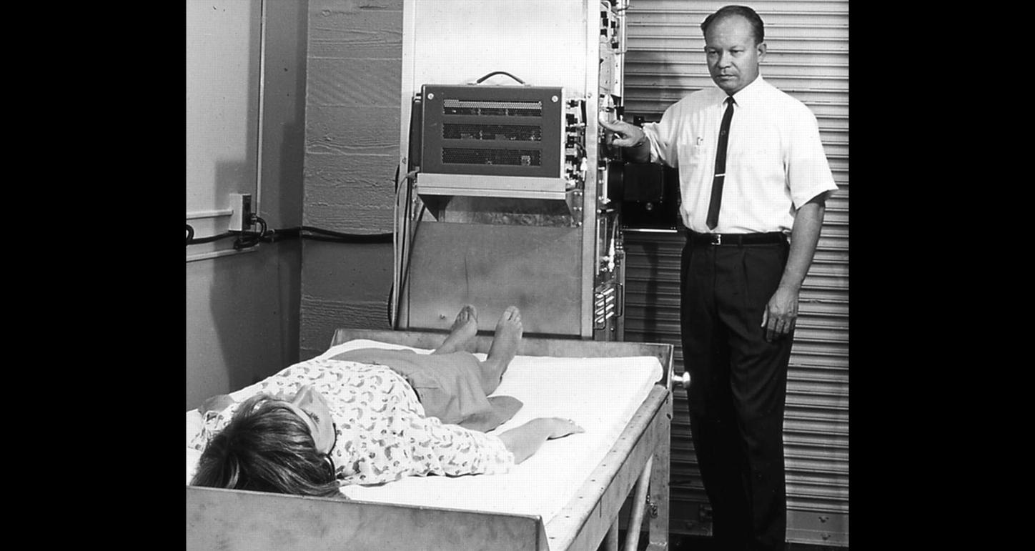
Hal O. Anger, the inventor of the gamma camera.
Known as Pho/Gamma 1, the device was a huge success for Nuclear Chicago, but it would still take around two decades for the gamma camera to become a standard instrument in molecular imaging. In the mid-1970s, many nuclear physicians still preferred rectilinear scanners for certain examinations. This was partly because they were used to them and partly because the devices also produced meaningful diagnostic results. Despite this, Nuclear Chicago increasingly focused its research funding on the development of gamma camera scanners. The company developed numerous major milestones on the path toward modern systems. It operated independently until the 1970s and then, following its acquisition in 1979, under the umbrella of the Siemens medical technology division. The basic principle developed by Hal O. Anger is still used in thousands of gamma cameras today. Anger’s invention defined the subsequent development of modern techniques such as single-photon emission computed tomography (SPECT) — or, indeed, was instrumental in making their development possible in the first place. In the words of the then president of the Society of Nuclear Medicine, H. William Strauss: “It’s hard to say where nuclear medicine would be without Anger’s contributions.”
The Pho/Gamma from Nuclear Chicago in 1972.
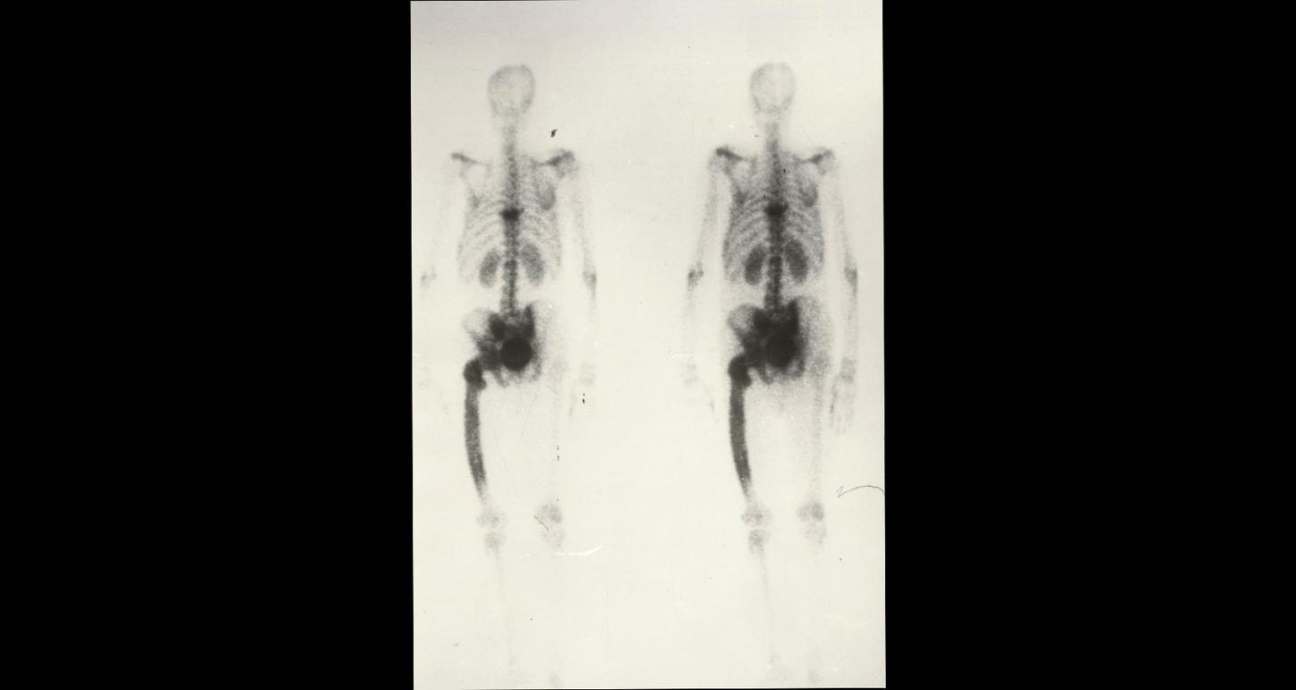
Bone scan with a gamma camera in 1972.
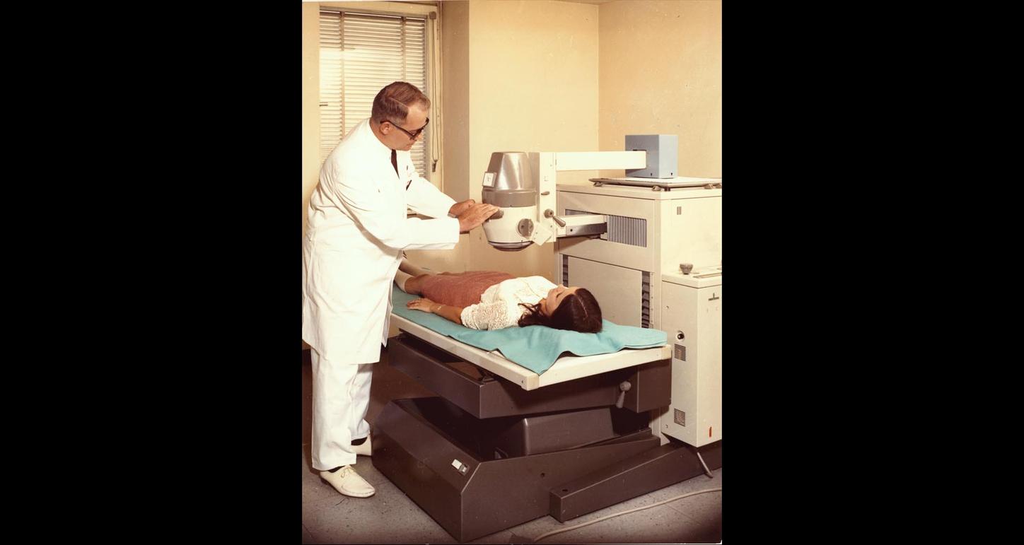
Until the mid-1970s, many physicians preferred rectilinear scanners such as the Scintimat 2 from Siemens.
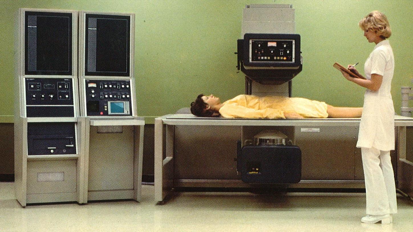
The first tomography system for molecular imaging, the PHO/CON Tomographic Multi-Plane Scanner from 1977.
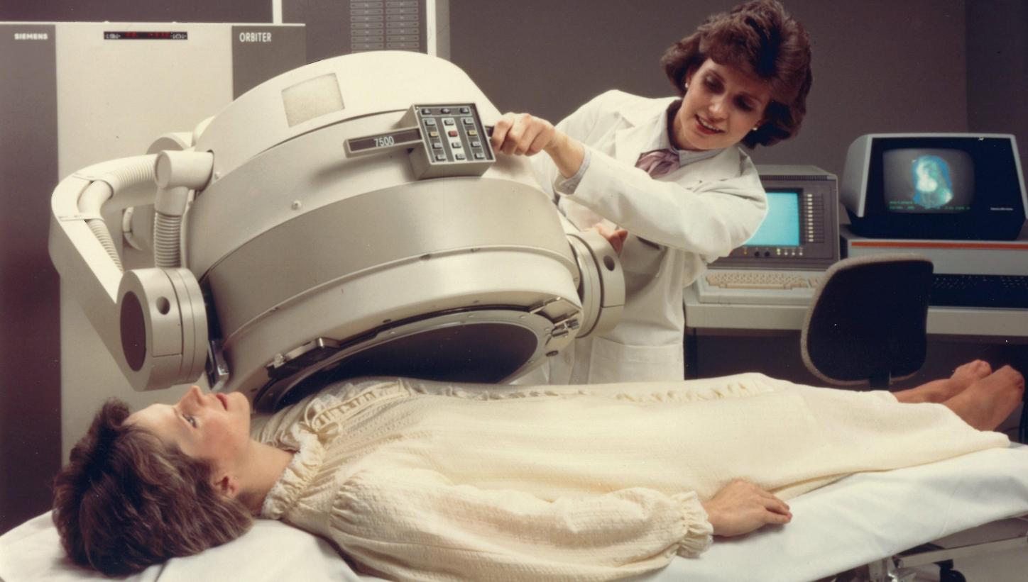
The Siemens Orbiter gamma camera became a best-seller in the 1980s.
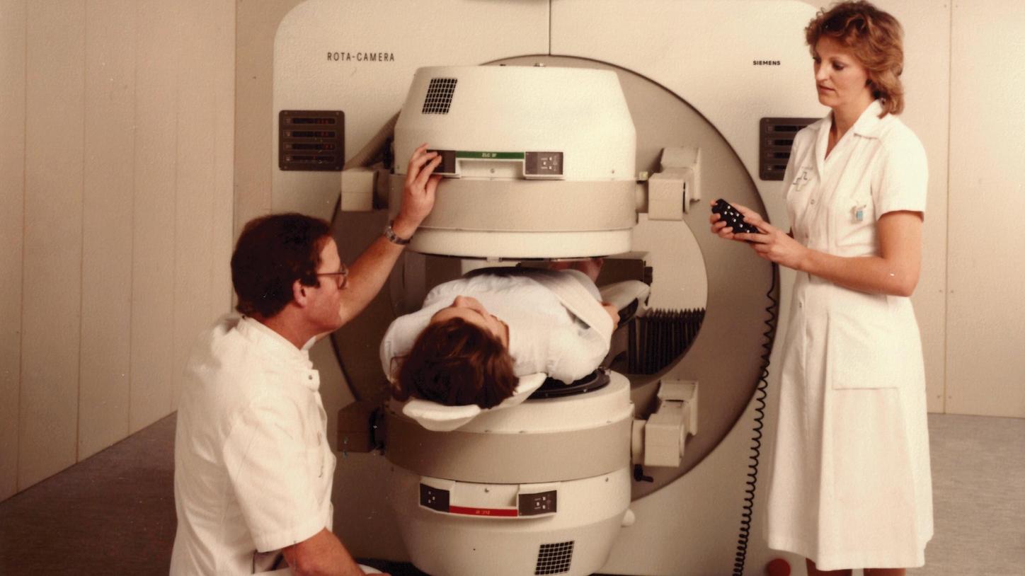
The Siemens ROTA camera, the world’s first commercial system with two rotating gamma cameras.
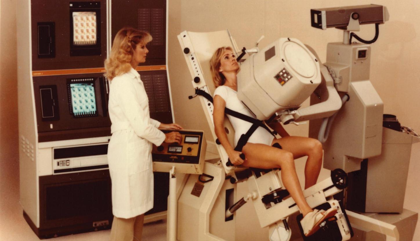
A heart scan with the Pho/Gamma LEM in 1983.
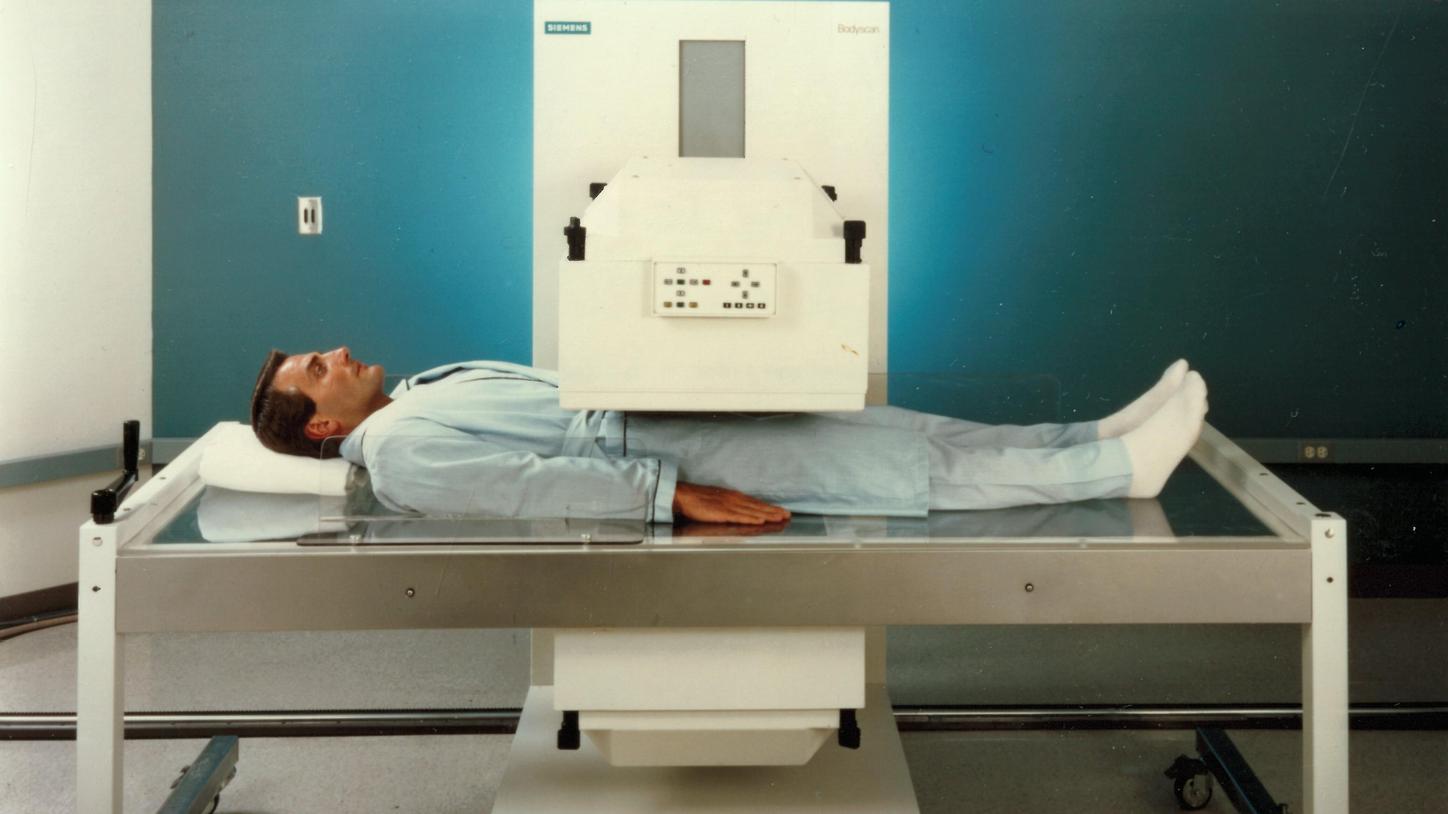
The Siemens BODYSCAN gamma camera from 1987, a system specially developed for whole-body scans.
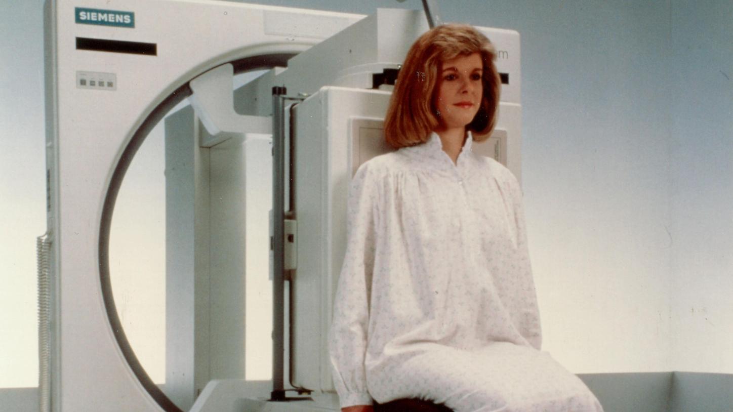
The Siemens DIACAM from 1990 was the first such device that could be configured for every common examination position.
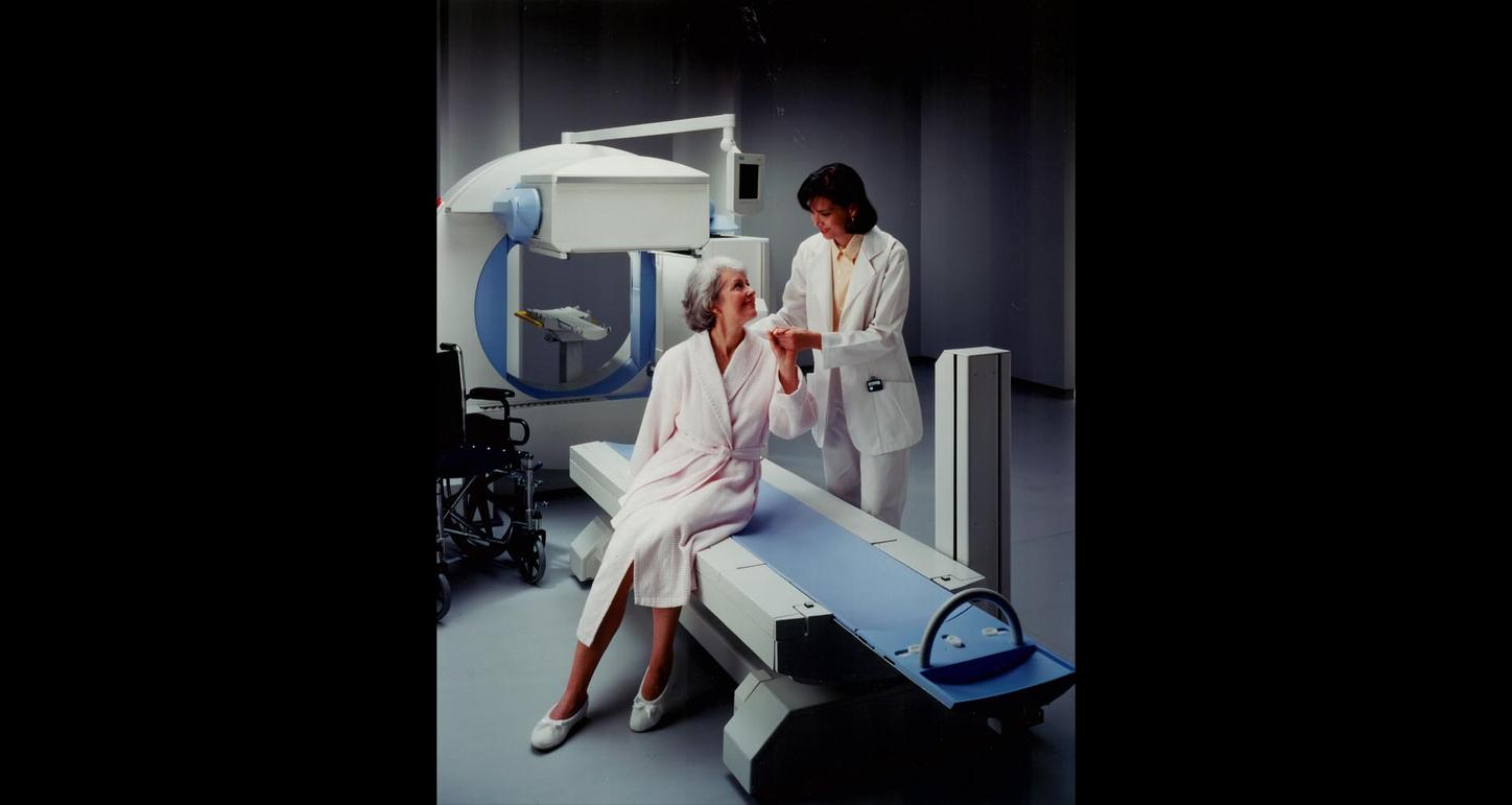
The flexible system architecture of the E.CAM from 1996 was already very similar to the basic framework for today’s SPECT systems from Siemens Healthineers.
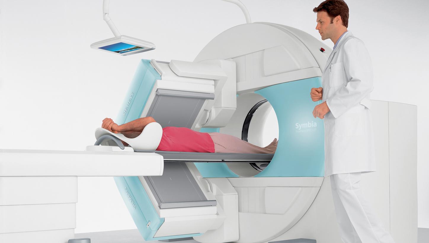
SPECT/CT TruePoint Symbia, the first generation of today’s Symbia systems in 2004.
The Pho/Gamma from Nuclear Chicago in 1972.
Technology journalist and author at the Siemens Healthineers Historical Institute